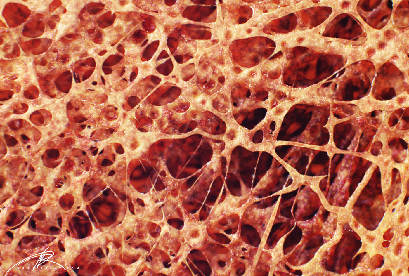
Bone Cross Section View - Vintage German Bone Structure Anatomical Model Femur ... : Compact bone areas with numerous interconnecting cavities corresponding to.
byAdmin•
0
Bone Cross Section View - Vintage German Bone Structure Anatomical Model Femur ... : Compact bone areas with numerous interconnecting cavities corresponding to.. Muscles of the rotator cuff labeled on a sagittal mr slice. Can you identify the primary and secondary haversian systems, central canals and bone lamellae? Cross section through middle metacarpal bones of vector. They are obtained by taking imaginary slices perpendicular to the main axis of organs, vessels, nerves, bones, soft tissue, or even the entire human body. The outside of a bone is covered in a thin layer of dense irregular connective tissue called the periosteum.
Cross section view of a human femur bone showing trabecula. The spongy and compact bone tissue in the cross section of a skull bone. (b) in this micrograph of the osteon, you can clearly see the concentric lamellae and central canals. Cross section of the head of the femur showing normal bone marrow versus. We can see there are two layers of compact bone here.
Axial skeleton. Cranium. Facial bones. Vertebral column ... from encyclopedia.lubopitko-bg.com This bone is located directly beneath the skin on the anterior aspect of the leg (top of the image). Generally speaking, it is very easy to recognize a cross section through the leg, mostly due to the tibia. Can you identify the primary and secondary haversian systems, central canals and bone lamellae? The male was sectioned at one millimeter intervals The osteon has blood vessels and bone cells, things vital for the survival of the bone. Cross section view of a human femur bone showing trabecula. Human bone cross section stock photos and images. The best selection of royalty free bone cross section vector art, graphics.
A cylinder of bone known as the vertebral body makes up the majority of the lumbar vertebrae's mass and bears most of the body's weight.
This photo shows a cross section through bone. Posteriorly the body is connected to a thin ring of bone known as the arch. Lamellar bone makes up the compact or cortical bone in the skeleton, such as the long bones of the legs and arms. Hope you enjoy and please. Label the membrane that lines the cavity and the membrane that covers the outside surface. Generally speaking, it is very easy to recognize a cross section through the leg, mostly due to the tibia. Online quiz to learn bone histology bone cross section. In addition, students can see the osteoid tissue, which is uncalcified matrix. The periosteum contains many strong collagen fibers that are used to firmly anchor tendons and muscles to the bone for movement. This bone is located directly beneath the skin on the anterior aspect of the leg (top of the image). When the bone section is viewed under transmission electron microscope, it is possible to see collagen that makes up most of the organic matrix. The osteon has blood vessels and bone cells, things vital for the survival of the bone. Cross section view of a human femur bone showing trabecula.
This photo shows a cross section through bone. Learn vocabulary, terms, and more with flashcards, games, and other study tools. Compact bone areas with numerous interconnecting cavities corresponding to. Mr of the shoulder mri of the shoulder : The osteon has blood vessels and bone cells, things vital for the survival of the bone.
"Bone Cross Section" for Radius Digital Science on Behance from mir-s3-cdn-cf.behance.net (b) in this micrograph of the osteon, you can clearly see the concentric lamellae and central canals. Most relevant best selling latest uploads. Cross section view of a human femur bone showing trabecula. Generally speaking, it is very easy to recognize a cross section through the leg, mostly due to the tibia. Smartdraw includes 1000s of professional healthcare and anatomy chart templates that you can modify and make your own. Hi all, i have uploaded a new medical animation tutorial. (b) in this micrograph of the osteon, you can see the concentric lamellae around the central canals. Learn vocabulary, terms, and more with flashcards, games, and other study tools.
Just behind the column of bones and discs is the sac which holds the nerves and fluid, called cerebral spinal fluid (csf).
Muscles of the rotator cuff labeled on a sagittal mr slice. The diagram above shows a longitudinal view of an osteon. Bone decalcification is the removal of the mineral component using an acid, leaving the bone soft and easy to cut. Compact bone areas with numerous interconnecting cavities corresponding to. Online quiz to learn bone histology bone cross section. The most important anatomical parts of the sagittal and axial images of a normal mri lumbar spine are pictured below. An mri of the shoulder of a healthy subject was performed in the 3 planes of space (coronal, axial, sagittal) commonly used in osteoarticular imaging, with two weightings to explore the musculoskeletal pathology of the. The osteon has blood vessels and bone cells, things vital for the survival of the bone. There are two ways to study bone histology. This file is licensed under the creative commons attribution 3.0 unported license.: Human bone cross section stock photos and images. Can you identify the concentric lamellae, central canal and the. This is a high power photo of a single haversian system.
Cross section of the leg through the soleus muscle: We can see there are two layers of compact bone here. This photo shows a cross section through bone. Two types of bone tissues in cross section of a long bone : This article lists a series of labeled imaging anatomy cases by system and modality.
Cross Section View Of A Human Femur Bone Showing Trabecula ... from l7.alamy.com We can see there are two layers of compact bone here. Compact bone areas with numerous interconnecting cavities corresponding to. By admin mei 10, 2021 they are obtained by taking imaginary slices perpendicular to the main axis of organs, vessels, nerves, bones, soft tissue, or even the entire human body. Hope you enjoy and please. Cross section view of a human femur bone showing trabecula. This is a high power photo of a single haversian system. As shown in figure 2. The osteon has blood vessels and bone cells, things vital for the survival of the bone.
Generally speaking, it is very easy to recognize a cross section through the leg, mostly due to the tibia.
Whereas a long bone has only one layer of compact bone (see fig 1). This article lists a series of labeled imaging anatomy cases by system and modality. Posteriorly the body is connected to a thin ring of bone known as the arch. This is a high power photo of a single haversian system. The osteon has blood vessels and bone cells, things vital for the survival of the bone. Hi all, i have uploaded a new medical animation tutorial. (b) in this micrograph of the osteon, you can clearly see the concentric lamellae and central canals. Human bone cross section stock photos and images. • a full cross section displays the cross section across the whole view, as shown in the two examples above. We can see there are two layers of compact bone here. An mri of the shoulder of a healthy subject was performed in the 3 planes of space (coronal, axial, sagittal) commonly used in osteoarticular imaging, with two weightings to explore the musculoskeletal pathology of the. Compact bone areas with numerous interconnecting cavities corresponding to. This is the same reason why the slightest touch hurts so much.
To start, select the structure on the model bone cross section. Cross section of the head of the femur showing normal bone marrow versus.